EMV for Windows
IMPORTANT NOTICE
This program requires QuickTime for Windows which has been discontinued. Until we make available a QuickTime-free upgrade, EMV v4 for Windows should be considered unsupported software.
EMV Windows is a robust, feature-rich and lightweight DICOM viewer, anonymizer and converter. The latest version gets and sends files to DICOM servers and PACS, opens virtually all DICOM images and DICOMDIR files from DICOM disks, and handles user objects, such as annotations and measurements.
If you're looking for a fast, friendly, powerful, multilingual, and overall trustworthy medical viewer, give EMV Windows a try.
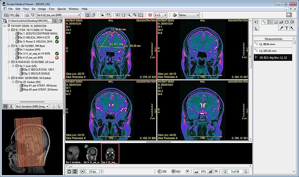
Click on the image above for more screenshots and use the arrows to navigate.
At a glance
General
- Multilingual interface: English, French, Italian, Spanish, Portuguese.
- Common image formats: JPEG, TIFF, etc (QuickTime supported formats)
- Full-screen viewing mode
- Printing to any connected system (non DICOM) printer
DICOM communications
- Query/Retrieve SCU: query PACS and DICOM servers to retrieve studies.
- Send SCU: send DICOM images to PACS and DICOM servers.
- Servers list: define all PACS and DICOM servers you usually work with.
Viewer
- Multiframe display (series, clips, sequences) in user-defined grid.
- User-controlled display of demographics and DICOM overlays.
- Alternative colorizations of grayscale and color images.
- Localizers: show CT/MR scout images with related slice locations.
- Handling of non-square pixels.
- Image calibration: define the real-world size of pixels.
- User-controlled action scope: act on the current frame or on the entire sequence.
- User-controlled marking: mark specific items for subsequent batch operation.
- Cine: time-vector support, user-defined frame rate, user-controlled autoplay.
User objects and measurements
- User objects: measurements, textual annotations, arrows
- Eraser tool for removal of information burnt into US clips.
- Density measurement in Hounsfield units, where applicable.
- Calibrated measurements on ultrasound regions, when present.
Special
- Series splitting of multislice or multiphase series
- Mask frame subtraction for XA clips
- Multi-Planar Reformation (MPR): reconstruction of 2D datasets typically from a stack of axial slices.
- Maximum and Minimum Intensity Projection (MIP/MinIP): volume-rendering techniques for enhancing contrast in CT datasets.
Batch conversions
- Export images: frame, sequence, series, study, patient, all patients, marked items.
- Anonymize: file, sequence, series, study, patient, all patients, marked files.
- Export DICOM data: file, sequence, series, study, patient, all patients, marked files.