| QMedical Command Reference |
|
DICOM images usually contain data encoded in the maximum resolution attainable by the modality that created them. For example, a grayscale image with 16 bits per pixel will have pixel values with a maximum dynamic range of 65536 = 216, e.g. values in the range 0 to 65535.
Visual representation of this many levels of gray is possible only on specialized monitors, whereas common PC displays can only show up to 256 levels of gray. As a result the original pixel values of an image (as they exist in the DICOM file) have to be squeezed into the range 0 to 255.
The worst case occurs when the original image has all 65536 levels, in which case 256 original levels will be replaced by 1 final level! You can imagine how huge (and unavoidable) loss of contrast can happen when displaying a 16 bit gray scale image on an 8 bit display.
Fortunately, the worst case does not occur so often because the tissues that are interesting to physicians usually occupy a small part of the available range of gray levels. So it is reasonable to try to map this interesting range to the available output range. This is exactly what is accomplished by using the VOI parameters and applying the VOI transformation.
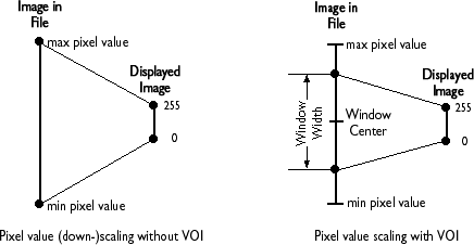
The VOI transformation specifies that only pixel values within 'Window Width' (WW) in the original image centered around the 'Window Center' (WC) will occupy the entire available output range, which is in most cases 0 to 255. Values lower than WC - WW/2 will be mapped to 0 and values greater than WC + WW/2 will be mapped to 255, i.e. values outside the range specified by the VOI parameters are to be clipped to the minimum and maximum output pixel values, respectively.
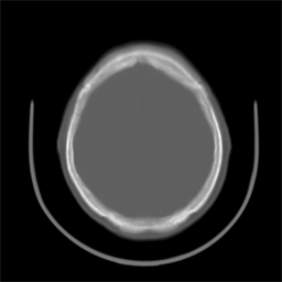
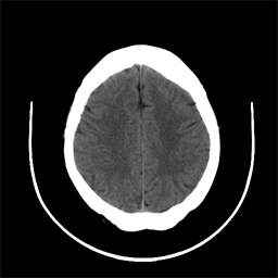
By appropriate manipulation of the VOI parameters one can dramatically affect the way image data are displayed; varying features of the image may become apparent or disappear as the WC and WW are modified. For example, consider the following two images; both were derived from the same DICOM image file of the cross section of a human skull. Note that in the left one, to which no VOI transformation has been applied, the brain area is almost uniformly gray and no structure is apparent. In the image on the right, to which the VOI tranfomration has been applied the internal structure of the brain tissue becomes strikingly apparent.
QMedical offers full support for DICOM VOI transformations. When an image is first assigned to a plug-in area, a default VOI transformation is applied to the image using the first existing duplet of VOI parameters available in the file, which are usually set to optimal values. If no such values exist in the file, default VOI parameters are set such as to result in an identity VOI transformation. The QMedical plug-in area includes end-user tools by which the VOI parameters can be adjusted interactively by tracking the mouse. VOI values are updated and displayed in real time in a tool-tip window near the upper-left corner of the image area.
Moreover, there are methods for retrieving and changing the VOI parameters programmaticaly. The VOI parameters respect the characteristics of the original image data such as their range (signed or unsigned; 8 or 16 bit) and photometric interpretation (MONOCHROME1 or MONOCHROME2 for gray scale images)
| QMed_GetVOILimits | Returns the minimum and maximum values of the VOI parameters |
| QMed_GetDefaultVOI | Returns the default values of the VOI parameters |
| QMed_SetCurrentVOI | Changes the current values of the VOI parameters |
| QMed_GetCurrentVOI | Returns the current values of the VOI parameters |